


| Technical Name | High-resolution, multiplex pancreatic imaging system | ||
|---|---|---|---|
| Project Operator | National Tsing Hua Univ | ||
| Project Host | 湯學成 | ||
| Summary | Our system integrates 3-D tissue scanning, high-resolution confocal imaging, and the gold-standard H&E histology to examine the pancreatic microstructure and neurovascular tissues with high definition. The 3-D fluorescence and 2-D H&E signals provide a side-by-side comparison and detailed spatial information of pancreas in a clinically related setting (e.g., diabetes), which otherwise cannot be achieved via standard histology. |
||
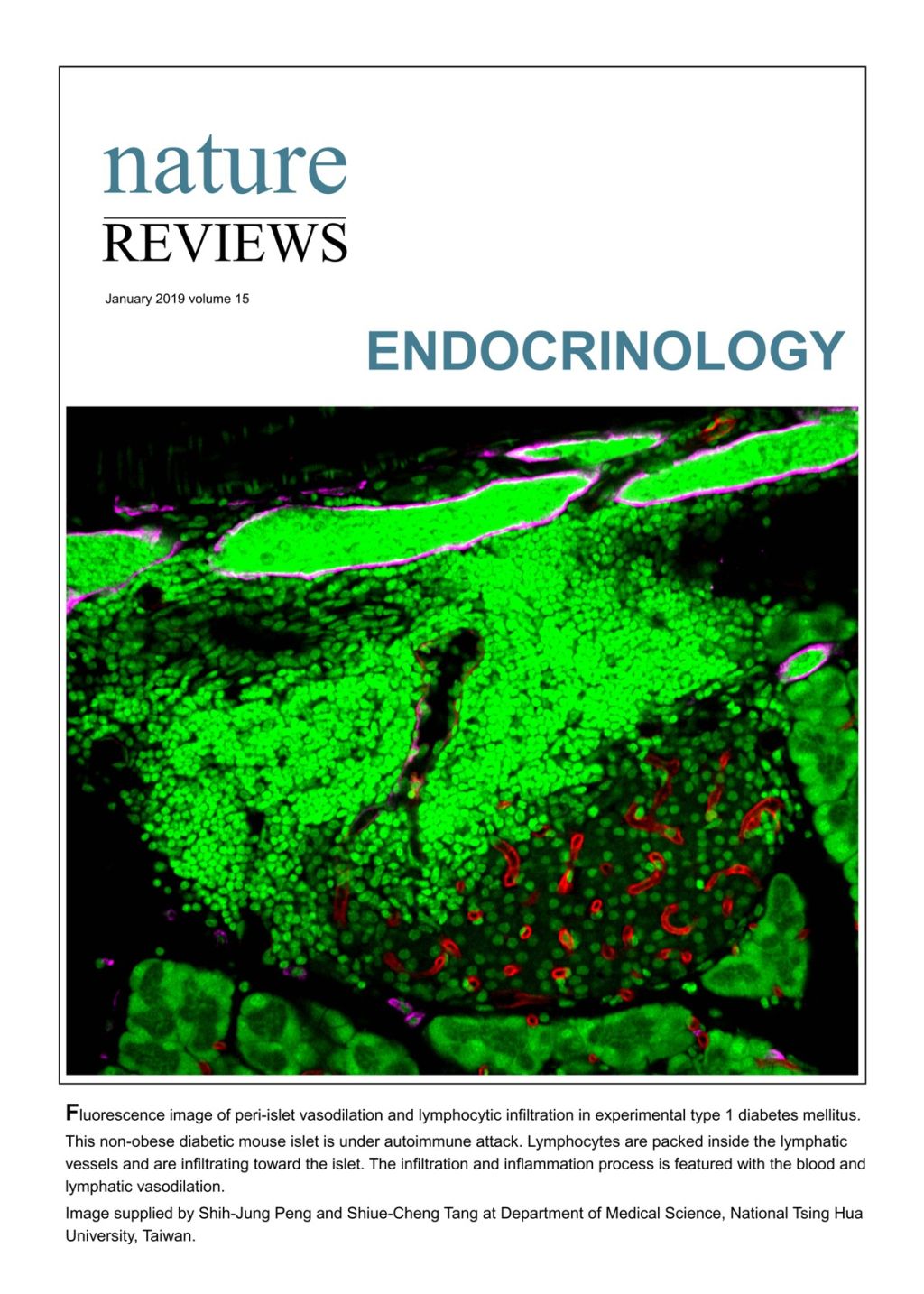
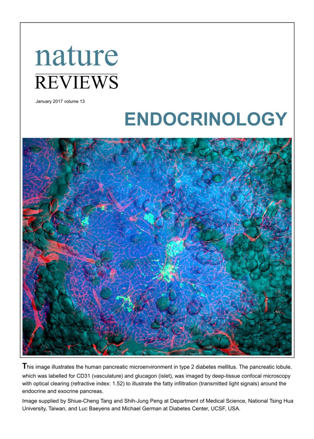
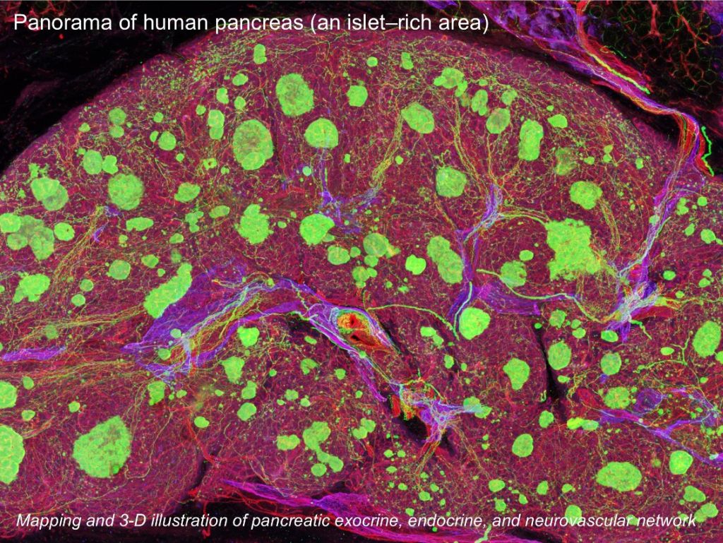
| Scientific Breakthrough | Our team develops 3-D histological tools and key knowledge in handling animal and human gut tissues for multi-dimensional and multiplex signal analysis. In pancreatic research we use the newly developed 3-D/2-D integrative histology to examine the human pancreases and tumor specimens, revealing the unknown details of pancreatic microenvironment in obesity, diabetes, and cancer lesion progression. |
||
| Industrial Applicability | The high-resolution multiplex imaging system can histologically analyze a variety of human and animal tissues, such as the pancreas, intestine, liver, kidney, and skin, in a global and integrated fashion. We are currently a member of MOST’s National Core Facility for Biopharmaceuticals, aiming to assist the academia and industry in 3-D/2-D tissue analysis with the state-of-the-art method to demonstrate Taiwan’s strength in biomedical imaging. |
||
| Keyword | high-resolution optical microscopy multiplex imaging system 3-D tissue scanning pathology 3-D fluorescence tissue imaging human pancreas diabetes pancreatic cancer neurovascular network lymphatic network | ||
- chienhj1026@gmail.com
other people also saw