Technical Name | Cancerous lesion identifying method via hyper-spectral imaging technique | ||
---|---|---|---|
Project Operator | National Chung Cheng University | ||
Project Host | |||
Summary | By the cancerous lesion identifying method via the hyper-spectral imaging technique in the present invention, the cancerous lesion images are digitized and the diagnosis rate of the clinicians may be efficiently increased to help the patients to start the treatments as soon as possible. |
||
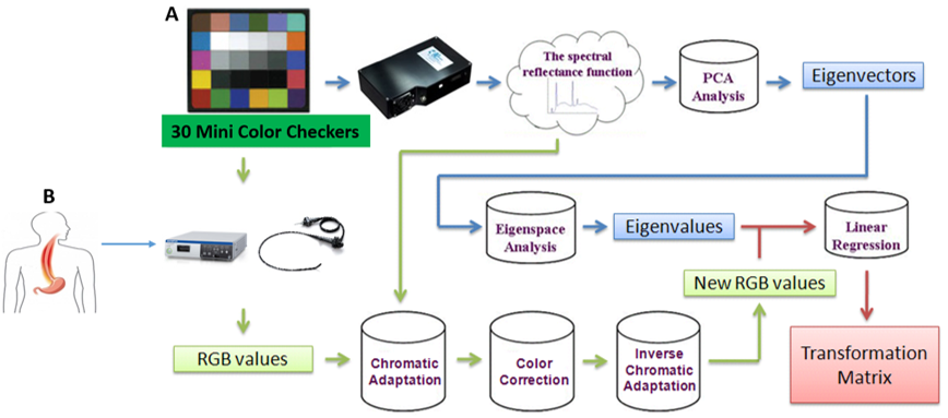
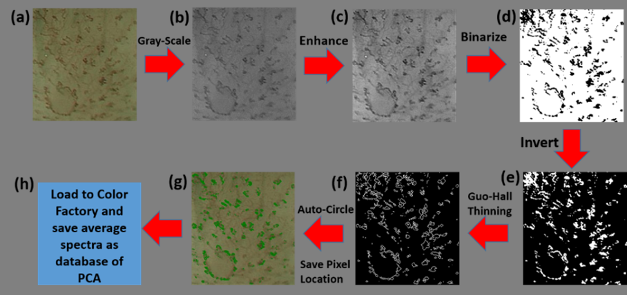
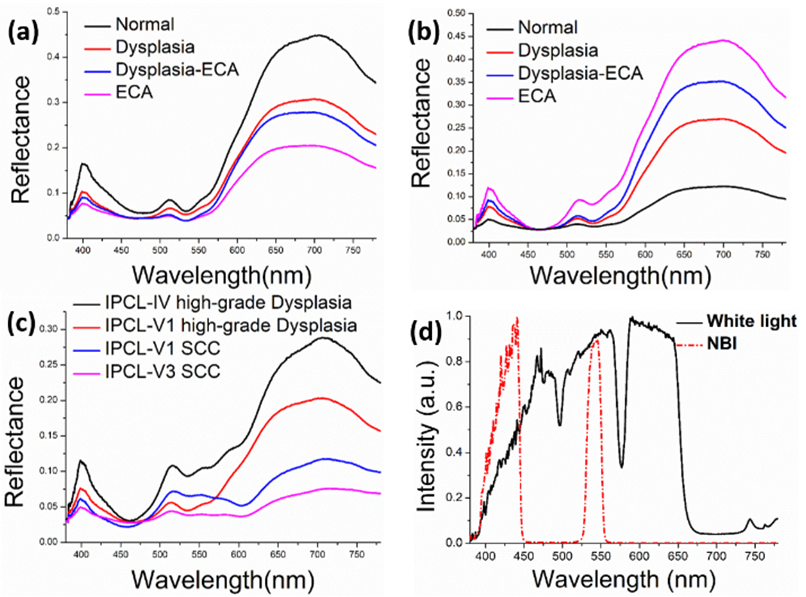
Scientific Breakthrough | In the detection of the esophageal cancer, the NBI is the current mainstream technology without spraying dye to include optical dye effect and the operation thereof is very easy. Only one button is to push to switch between the white light mode and the NBI mode so as to repeatedly observe. However, the detection is based on observing surface vessels, such as the changes in Intra-epithelial Papillary Capillary Loop (IPCL), much relying on the subjective determination of the clinicians. Therefore, if the hyper-spectral imaging technique can work with the principle component analysis, a new optical detection method may be provided. |
||
Industrial Applicability | 光譜影像在癌症研究上扮演重要角色,但於我國發展仍處於萌芽階段,原因在於難以取得光譜影像,國科會精密儀器發展中心所發展光譜影像儀系統,可協助國內研究團隊於光譜領域的研究,近來在國外已有多篇論文討論光譜影像於上述領域的研究,很明顯各項預測分析很能符合現況需求,就產業經濟而言,未來光譜影像儀走向模組化後,可植入精密儀器系統,取得各空間完整光譜資料,這對癌症在反應前後的空間分布分析有很大的幫助。 |
||
Keyword | |||
Notes |