


| Technical Name | Personal brain structure displaying device having intracranial electrodes and its displaying method | ||
|---|---|---|---|
| Project Operator | Taipei Medical University | ||
| Project Host | 彭徐鈞 | ||
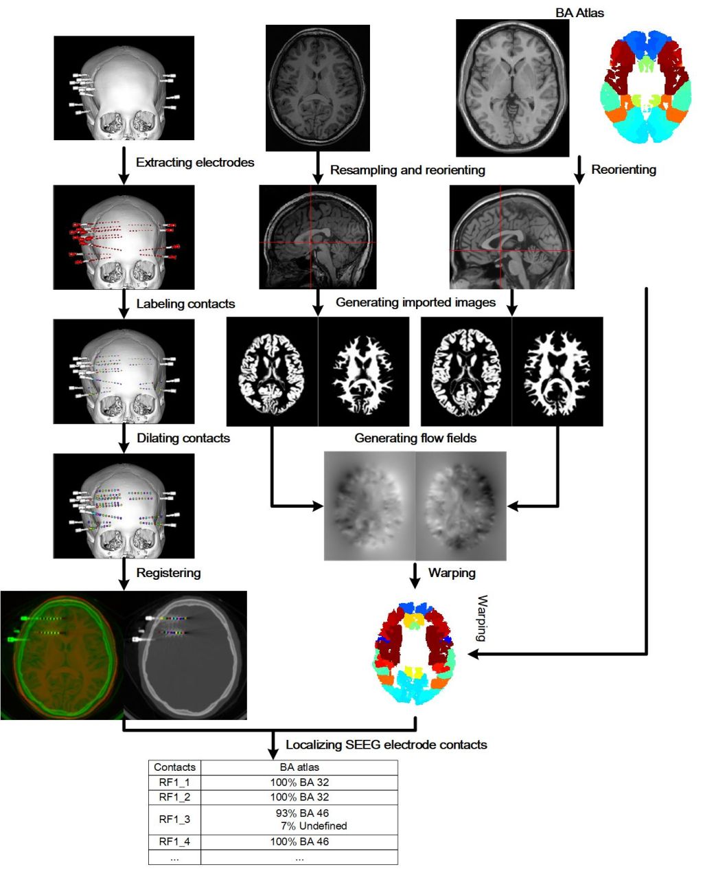
| Summary | The methodological procedure was developed to avoid the requirement for highly specialized medical resources and reduce time to identify the neuroanatomical location of epileptogenic zones. The co-labeling algorithm uses pre-implant magnetic resonance imaging and post-implant computed tomography fusion for the localization of epileptogenic zones and its relation with automatic labeled surrounding cortex and fiber bundle. |
||
| Scientific Breakthrough | The technique via different universal brain atlases for localizing intracranial electrodes in an epilepsy patient, and more details for image processing are provided, particularly in localizing electrode contacts in a CT image, and in acquiring a personalized brain atlas. The technique can reconstruct a 3D model of cortical surfaces based on the registered and reoriented pre-electrode-implantation MR image. |
||
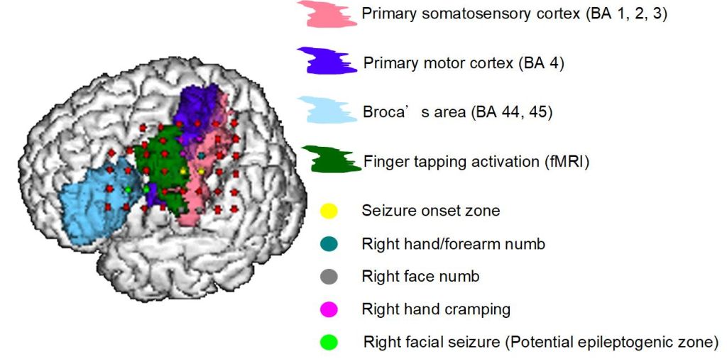
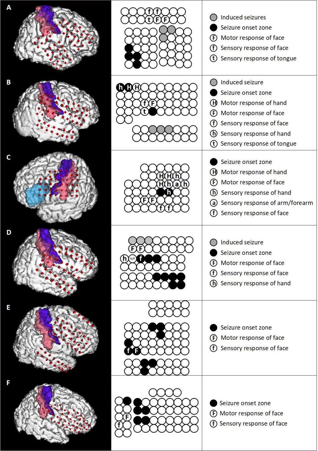
| Industrial Applicability | The technique can be applied in localizing electrodes implanted intracranially, e.g., SEEG electrodes or DBS electrodes. Intracranial electrodes can be used for localizing a specific epileptic discharge site in a subject's brain. DBS electrodes can be used in patients requiring deep brain stimulating or requiring electrode placed in the brains, such as patients with Parkinson's disease, tremor, etc. |
||
| Keyword | Intracranial electrode localization Electrocorticography Epilepsy surgery Brain mapping Cortical electrical stimulation Precision Medicine Neuromodulation Prognosis evaluation Electrocauterization Stereotactic electroencephalography | ||
- sjpeng2019@tmu.edu.tw
other people also saw