


| Technical Name | Manufacturing Lifelike, Fully Transparent, and Elastic Cerebral Circle of Willis Vascular System as An Education Simulator for Neurosurgeons. | ||
|---|---|---|---|
| Project Operator | National Taiwan University of Science and Technology | ||
| Project Host | 陳品銓 | ||
| Summary | Neurosurgeons require solid expertise and substantial practical experience to perform difficult surgeries and avoid critical situations. Unfortunately, medical students have limited opportunities to develop skills in the operating room. A key aspect of surgical training involves the use of cadavers to give trainees practical experience navigating the structure of the brain under the trained eye of senior surgeons. However, cadavers are expensive and can be difficult to obtain, and the vascular morphology of cadavers often differs from that of actual patients. Recent advances in virtual reality (VR) and 3D printing technology have opened the door to the medical simulators; however, such devices remain expensive, the imaging modalities differ from those encountered in actual 3D spaces, and they lack the tactile feedback. Our biomodel has substantially closed the gap between the medical simulators and the situations of actual patients in a real-world surgical environment. |
||
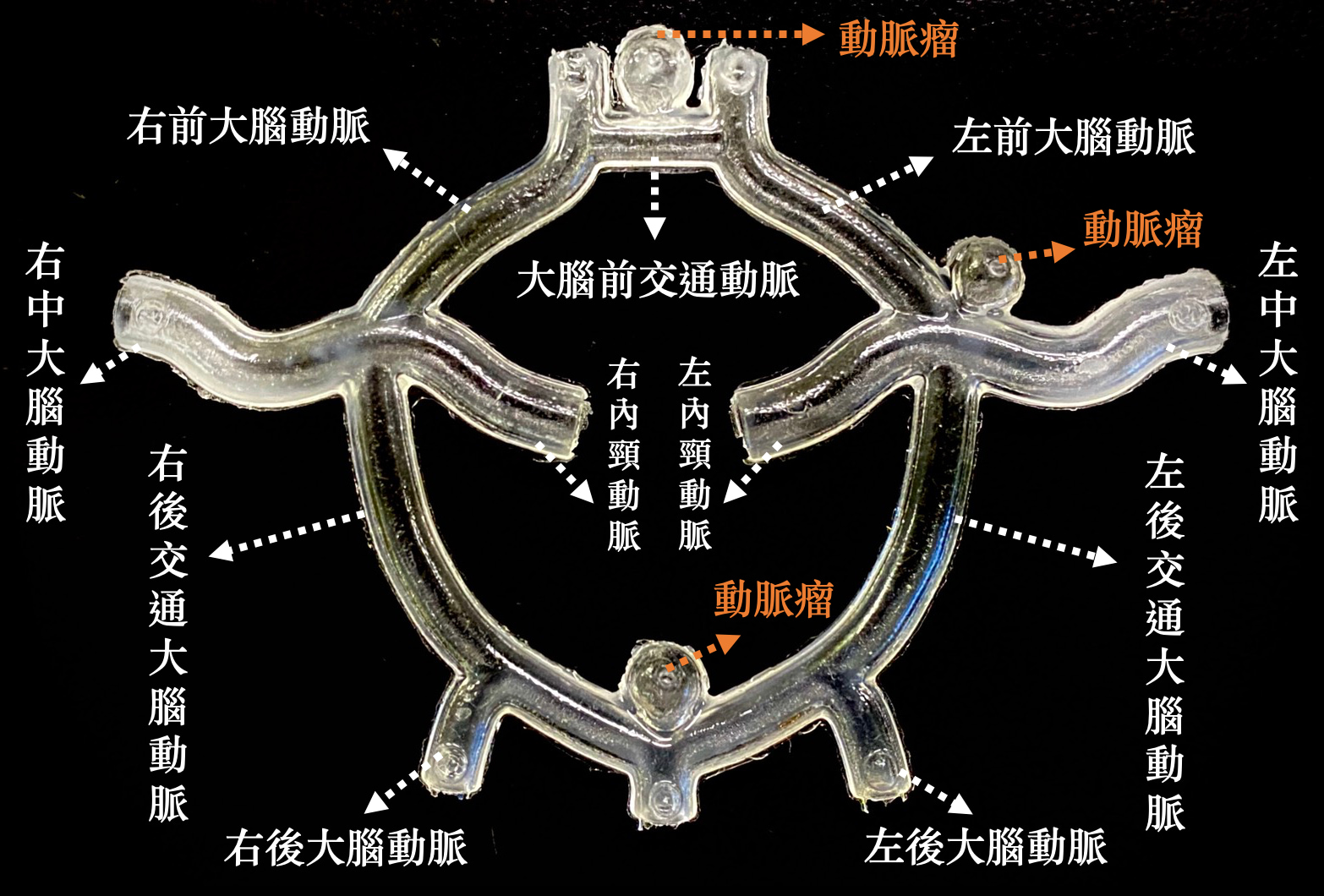
| Scientific Breakthrough | Until now, researchers have lacked the process technologies to create complete vascular models (e.g., the Circle of Willis) for experiments. The dimensional inaccuracy, opaqueness, and/or inelasticity of 3D printed medical models hardly can be used as lifelike biomodels. Our technology allow control over the thickness of vascular walls, and makes it possible to create a hollow, highly complex, and entirely transparent model of the vascular system within the brain. In the proposed model, we achieved a 3.2% coefficient of variation in vascular wall thickness as well as a 2% coefficient of variation in vessel diameter, while the elasticity coefficient of the vascular walls was 1.8 Mpa, which is quite matching the characteristics of real blood vessels (1.7 Mpa). |
||
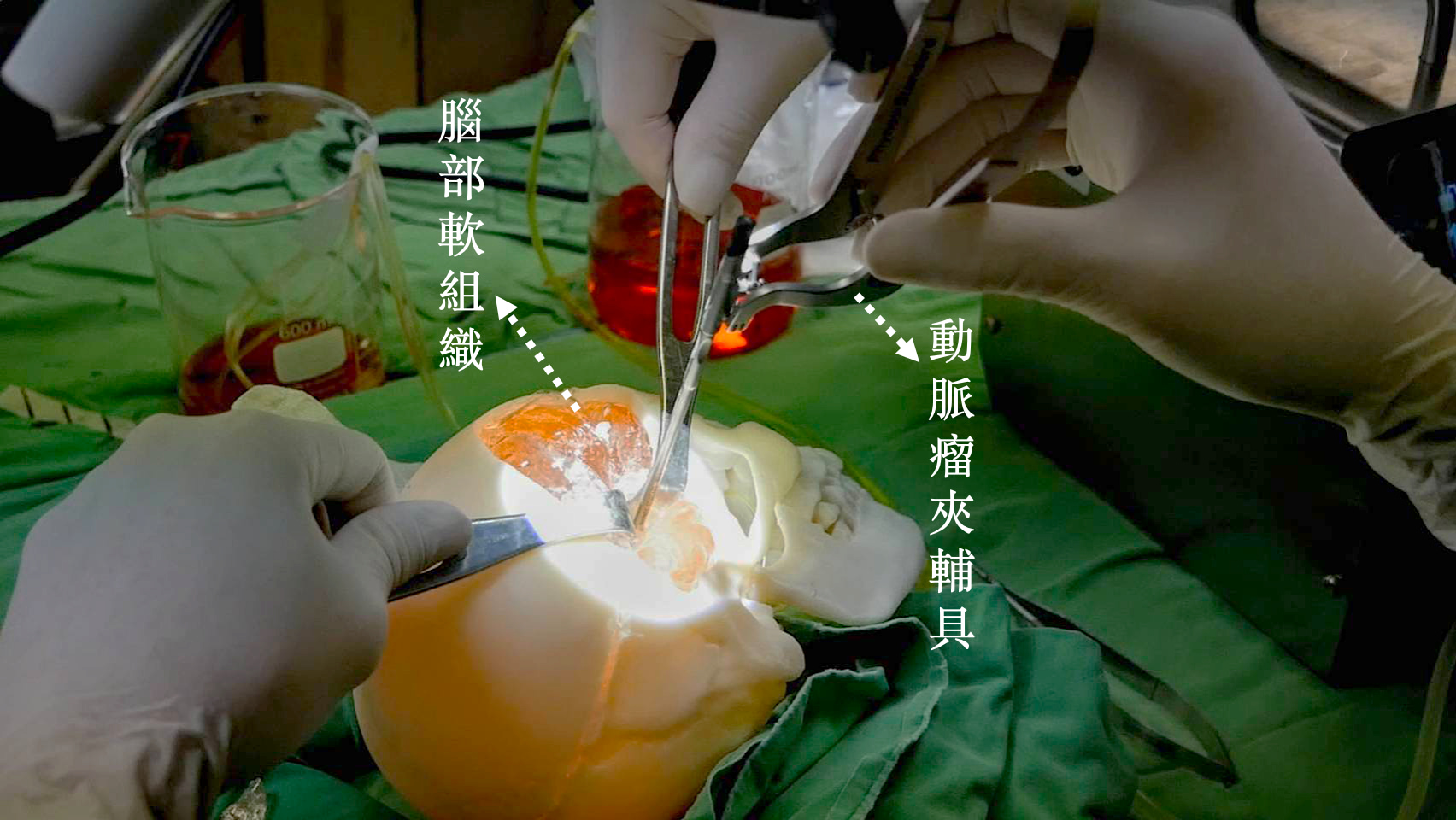
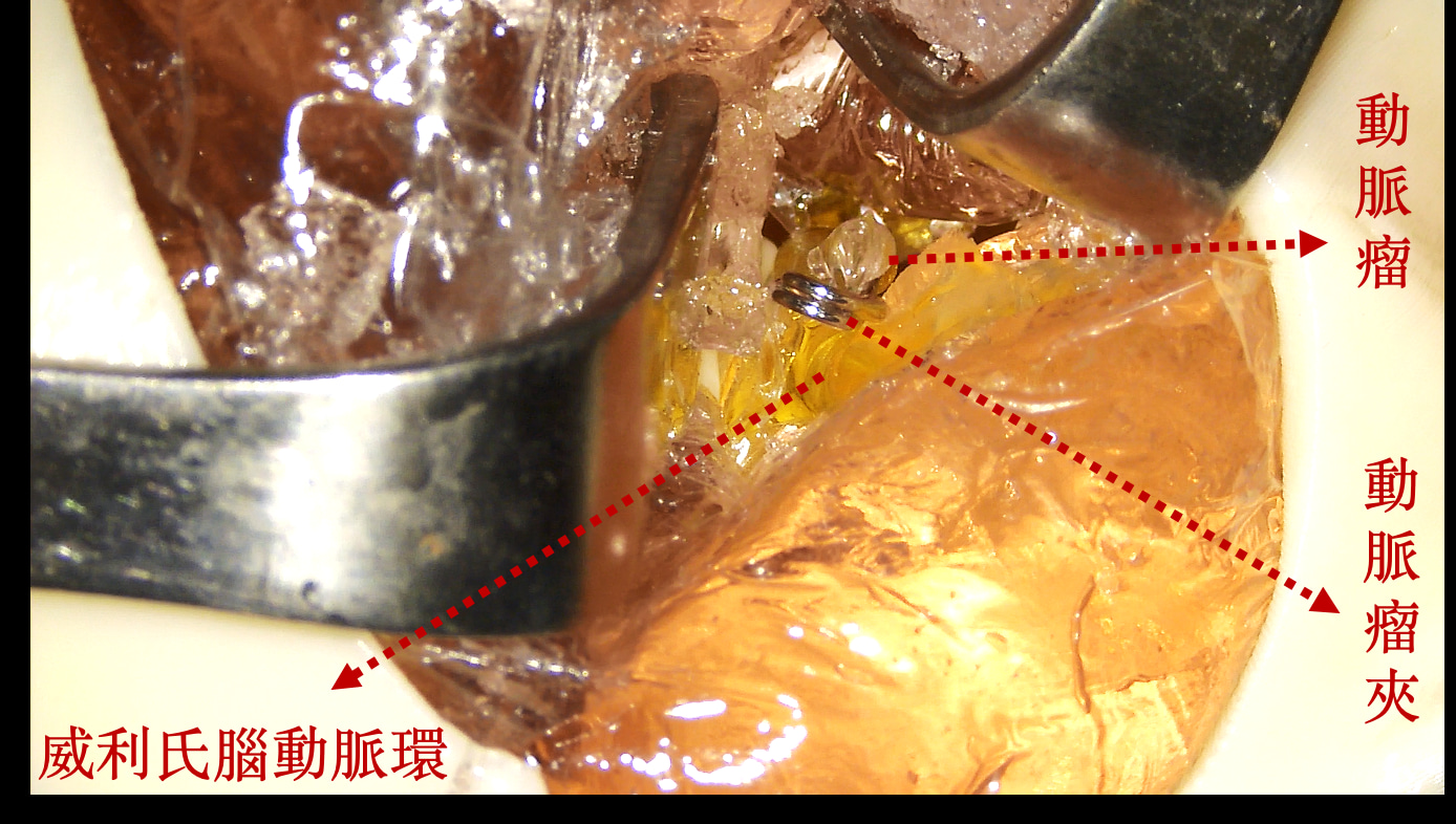
| Industrial Applicability | This biomodel has substantially closed the gap between the medical simulators and the situations in a real-world surgical environment. It is applicable in a broad range of fields. (1) The biomodel could be applied in the conversion of medical images of a particular patient into 3D models, for use by surgeons in performing pre-operative simulations aimed at experiencing difficult surgeries and increasing success rates. (2) The biomodel could be used as visual aids for surgeons explaining procedures to patients. (3) The biomodel could be used in fundamental research, such as exploring the reasons of the formation and rupturing of aneurysms. The cost of a biomodel is roughly NTD 20,000, which is significantly less than the cost of acquiring a single cadaver or installing a VR system. |
||
| Keyword | Lifelike Vascular System Cerebral Aneurysm Biomodel Neurosurgery Medical Education Training 3D Printing Biomodel Mold Design Casting of Elastic Materials Lifelike Aneurysm Lifelike Blood Vessel | ||
- M10703117@mail.ntust.edu.tw
other people also saw