


| Technical Name | Oral Image Analysis System and Method | ||
|---|---|---|---|
| Project Operator | National Cheng Kung University | ||
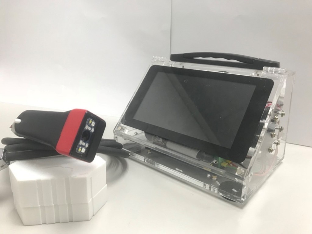
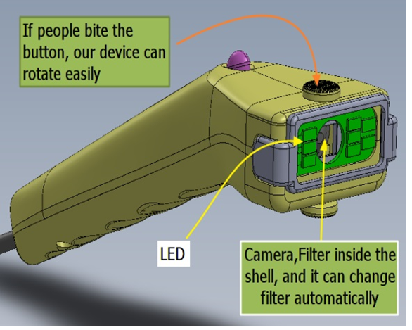
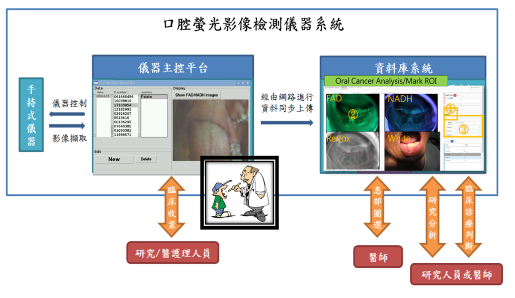
| Summary | The hand held device captures dual-spectrum autofluorescent images and use devised image analysis methods for extracting features, which can best distinguish cancer from normal lesions. Embedded inside the system is also a deep learning model which can automatic detect the suspect regions from buccal areas. |
||
| Scientific Breakthrough | 1: The design of the dual spectrum autofluorescent image analyzer is based on cancer optical properties to select the spectrum images which can reveal the best distinguishing between tumor and normal tissues. |
||
| Industrial Applicability | 鱗狀上皮細胞病變或附屬器官細胞惡性腫瘤檢驗等等相關檢測或輔助治療應用。 |
||
other people also saw